

Members of the project


Introduction
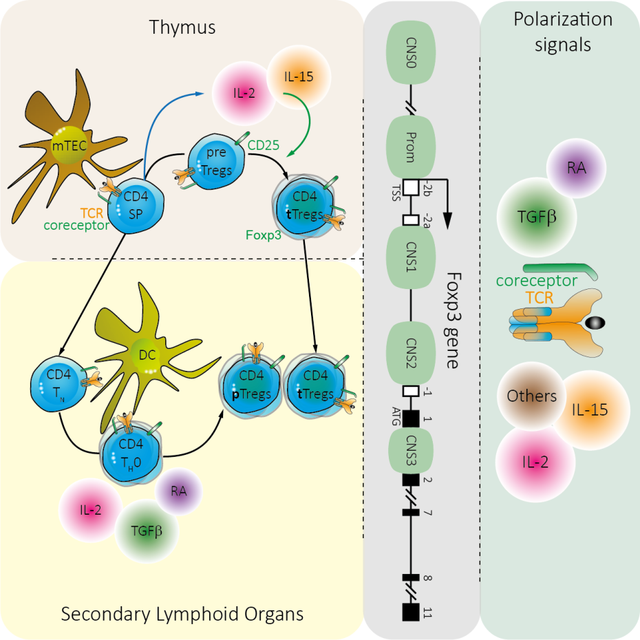
Regulatory T cells (Treg cells) are the main mediators of peripheral tolerance under physiological settings. In the periphery, Treg cells encompass cells that have been naturally produced within the thymus, called thymic Treg cells (tTreg cells), and cells with similar phenotype and functions that have differentiated from naïve CD4 T cells following antigen recognition in secondary lymphoid organs, called peripheral Treg cells (pTreg cells).
Although they share a similar phenotype (characterized in particular by the expression of the transcription factor Foxp3) and equivalent functions, tTregs and pTregs are only partially redundant in inhibiting immune responses. The complementary role of tTreg and pTreg cells, demonstrated for instance in the prevention of autoimmune diseases could be explained in part by their distinct TCR repertoires. Indeed, whereas tTreg cells, generated in response to interactions with the Self (Self peptide complex - Self major histocompatibility complex (MHC)) during their selection in the thymic medulla, display a repertoire biased towards the Self, that of pTreg cells is potentially broader. Importantly, the mechanisms underlying the generation of these 2 subpopulations of Treg cells also differ. Indeed, in contrast to tTreg cells, the emergence of pTreg cells from naïve CD4 T cells is controlled by TGFβ. TGFβ signaling pathways and response elements within the Foxp3 gene (CNS1) thus drive pTregs differentiation without affecting tTregs generation.
While defects in the development of thymic Tregs lead to immune dysregulation and life-threatening systemic autoimmune manifestations, the role of pTregs has so far only been demonstrated in the prevention of inflammation of the intestine or lung mucosa and in fetomaternal tolerance. It remains to be determined whether pTregs participate in immune regulation in other homeostatic or pathological contexts.
Aim #1
Deciphering the TGFβ-dependent signaling pathways leading to the generation of peripheral regulatory T cells
While TCR and IL-2 signaling pathways dictate tTreg cell generation from thymic precursors (preTreg cells), TGFβ signaling is crucial for the differentiation of naïve CD4 T cells into pTreg cells in vitro and in vivo. TGFβ binds to a hetero-tetrameric receptor complex composed of TβRI and TβRII that triggers the canonical SMAD pathway as well as numerous other signaling modalities, including MAPK, AP-1, PI3K and NFκB pathways. In the canonical SMAD pathway, SMAD2 or SMAD3 (SMAD2/3), phosphorylated by TβRI, form a complex with SMAD4 and translocate into the nucleus for transcriptional regulation. Foxp3 gene expression is controlled by a core promoter and at least 4 enhancers (conserved noncoding sequences, CNS0 and 1-3). Each regulatory element contains a variety of transcription factor binding sites which regulate foxp3 promoter activity. CNS1 is located between exons -2b and -1 of the Foxp3 gene and besides transcription factor binding sites for Rel-NFAT, CREB, RAR/RXR and AP-1 harbors a consensus sequence for Smad3. Consistently, mice with a targeted deletion of the Foxp3 CNS1 enhancer (CNS1KO mice), or deficient for Smad4 (Smad4KO) or Smad2 and Smad3 (Smad2/3DKO) have an impaired commitment of naive T cells to the pTreg cell lineage in vitro.
However, this impaired ability of naïve CD4 T cells from Smad4KO, Smad2/3DKO or CNS1KO mice to commit into pTreg cells is only partial. Indeed, in vitro Treg cell polarization assays in response to TGFβ reveal a 50% decrease in the proportion of Foxp3-expressing cells arising from naïve CD4 T cells from KO mice when compared with WT mice. Altogether, these results suggest that half of the TGFβ dependent conversion of naïve CD4 T cells into Treg cells is Smad- and CNS1-independent.
Using newly generated deficient mice, we now plan to decipher TGFβ-dependent Smad-independent signaling pathways leading to pTreg cell generation in vitro and in vivo.
Aim #2
Role of neonatal regulatory T cells in the prevention of autoimmune diseases
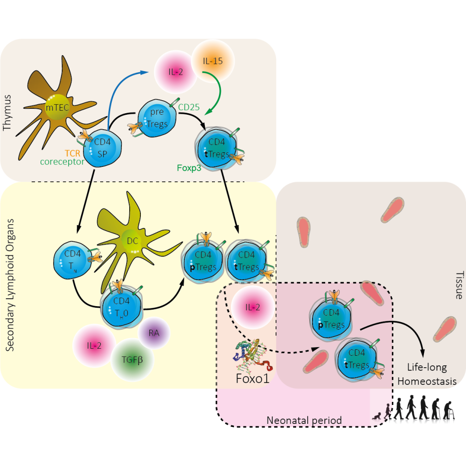
The incidence of autoimmune diseases (AIDs) has recently increased sharply in our societies. Epidemiological data suggest that this rise is partly attributable to changes in the exposure of young children to microorganisms. These would play an ambivalent role, as the commensal microbiota would be protective while certain pathogens could increase susceptibility to the development of these diseases with age. These observations could be explained by the effect of these microorganisms on Foxp3+ regulatory T lymphocytes (Tregs), which play a key role in the prevention of AIDs. Indeed, certain Tregs generated during the neonatal period (nnTregs), which appear to persist into adulthood and thereby control these diseases, may have their development positively modulated by the microbiota and negatively modulated by certain pathogens. At the molecular level, the transcription factor Foxo1 — which controls the sensitivity of these cells to IL-2, a cytokine critical for their expansion during this period — would play an essential role in the development of nnTregs. The overarching objective of this project is to investigate the impact of nnTregs on the control of AIDs in mice, with particular attention to the role of Foxo1 in their generation and to the means by which nnTregs can be modulated by microorganisms. First, we will determine whether modulation of nnTregs by targeting Foxo1 or IL-2 — using blocking antibodies or specific inhibitors — genuinely plays a major role in the prevention of AIDs in adults. We will then dissect the molecular mechanisms governed by Foxo1 in nnTreg development. Finally, we will seek to demonstrate whether inflammation triggered by certain pathogens during the neonatal period has a detrimental effect on nnTregs and on the prevention of AIDs. Indeed, recent data from the laboratory indicate that inflammation downregulates Foxo1 expression. This project should provide a better understanding of the reasons behind the rising incidence of AIDs and the role of nnTregs in their control, and could open up new therapeutic perspectives.
Publications
Fundings