


The MT1 Melatonin Receptor Switches Sides at Dawn: A New Molecular Mechanism Unveiled
Melatonin is a neurohormone primarily secreted rhythmically by the pineal gland, with production peaking during the night and being virtually absent during the day, thereby serving as a key signal for circadian and seasonal regulation in mammals. Its biological effects are mediated by two G protein-coupled receptors (GPCRs), MT₁ and MT₂ , which are proteins featuring seven transmembrane domains expressed in numerous tissues, notably the brain, retina, and pituitary gland. These two receptors exhibit high affinity for melatonin and are classically described as being coupled to Gi proteins, a family of heterotrimeric proteins whose activation leads to the inhibition of adenylyl cyclase and, consequently, to a decrease in the production of cyclic AMP (cAMP), a second messenger molecule essential to numerous intracellular signaling pathways.
In this study, the authors demonstrate that the MT₁ receptor is capable of coupling not only to Gi proteins, as expected, but also to Gs proteins: heterotrimeric proteins that, unlike Gi, stimulate adenylyl cyclase, thereby increasing intracellular cAMP levels and activating downstream effectors such as protein kinase A (PKA) and the transcription factor CREB (cAMP Response Element-binding protein).
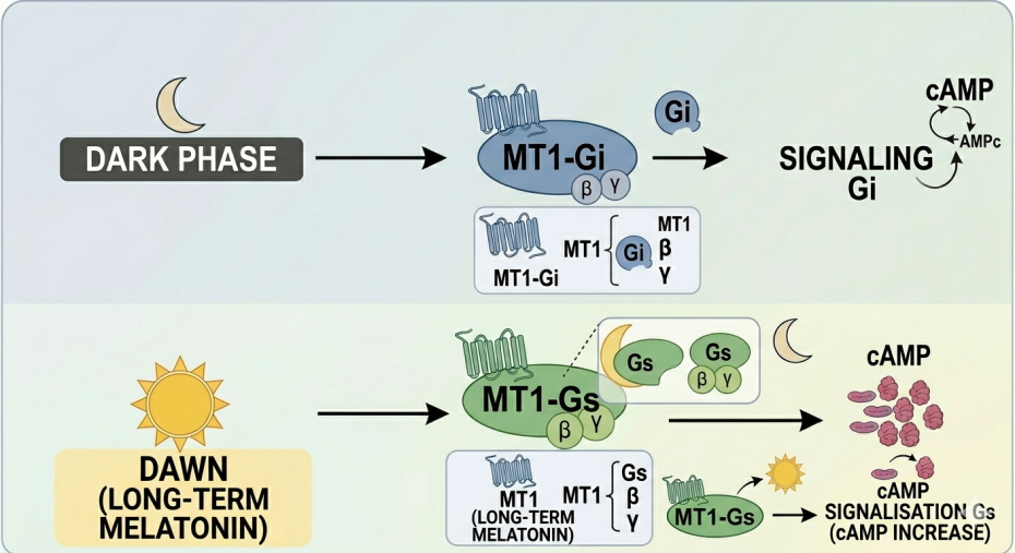
The coupling of MT₁ to Gs proteins occurs in a dose- and time-dependent manner: *in vitro*, in HEK293 cells, at low melatonin concentrations, MT₁ inhibits cAMP production via Gi. Conversely, at high doses, or following a prolonged 16-hour exposure mimicking conditions at dawn (the end of the nocturnal phase), MT₁ stimulates cAMP production via Gs; this effect has been confirmed *in vivo* in the mouse *pars tuberalis* (a bundle of axons projecting from the hypothalamus into the pituitary gland), using CREB phosphorylation as an activation marker.
To understand the molecular basis of this atypical coupling, the authors resolved the cryo-EM (cryogenic electron microscopy) structure of the MT₁-miniGs complex at 3.0 Å, thereby revealing a binding mode fundamentally different from that of the MT₁-Gi complex: the α5 helix of Gs inserts superficially on the side of the ICL2/TM3 protein domains, whereas that of Gi penetrates more deeply toward the TM5/TM6 domains, involving key residues Tyr128 and Leu133, mutations of which abolish the stimulatory response. Subsequent MT₁/MT₂ chimera experiments demonstrated that grafting the TM5-ICL3-TM6 region from MT₁ onto MT₂ is sufficient to confer Gs coupling. Only the minimal sequence comprising the first 12 amino acids of the MT₁ ICL3 domain (VRQRVKPDRKPK) appears necessary to confer this Gs coupling, notably via residue Val217, which stabilizes the interface with Gs.
In conclusion, from a structural perspective, this work reveals that a single receptor can engage two G proteins with opposing roles via radically distinct binding modes, with ICL3 emerging as a central and unexpected player in the coupling to Gs. Physiologically, this Gi/Gs switch constitutes an elegant mechanism through which MT₁ could encode not only the presence of melatonin but also its duration of action, thereby translating circadian temporal information into differentiated intracellular signals within the pars tuberalis. These results open up new therapeutic avenues, suggesting that selectively targeting one or the other of MT₁’s coupling modes could enable the fine-tuning of circadian transitions, with potential implications for the treatment of biological rhythm disorders, sleep disturbances, and pathologies associated with dysregulated melatoninergic signaling.
Figure legend: At night, the MT1 receptor is preferentially coupled to the inhibitory G protein (Gi), suppressing cAMP production. At dawn, prolonged exposure to melatonin triggers a switch to the Gs protein, thereby activating the cAMP pathway.
To know more about:
Nat Commun. 2026 May 21. doi: 10.1038/s41467-026-73555-6. Online ahead of print. PMID: 42168230


