Thick tissue cutting
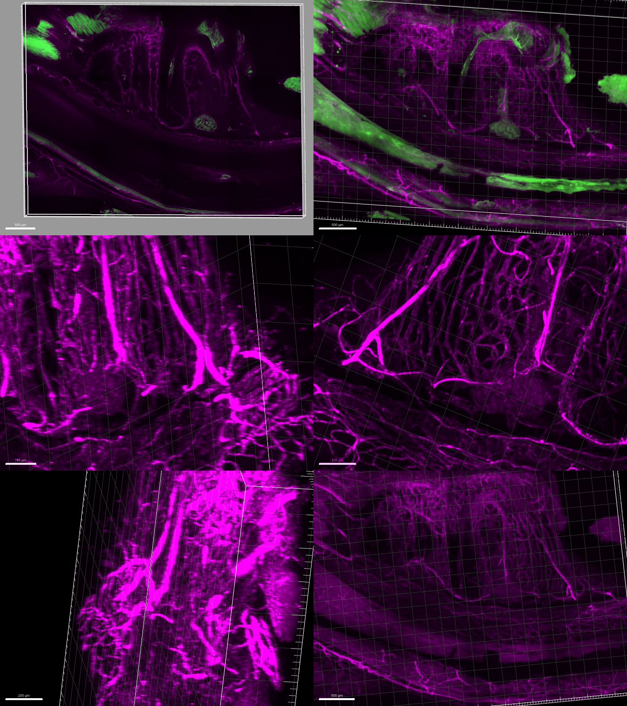
From fresh or fixed murine or human samples, the sections are made with a vibratome (VT 1200S, Leica) located in BSL2. The tissue is embedded in low gelling agarose which will serve as a support and facilitate the sectioning. The sections (100µm to 500µm) are collected and stored before being used for clarification or in vivo imaging
Some of our microscopes are in L2 containment, they are equipped with thermostatically controlled chambers, with the necessary equipment to perform perfusion and time-lapse, in adequate temperature and gas exchange conditions, allowing for example to perform FRET, calcium imaging.
A service request form is available on Openiris https://openiris.io/
Tissue clearing
Tissue clearing consists in making optically opaque samples transparent and allowing a better in-depth analysis of their structure. After immunolabeling of the sample (minimum 1 week), the tissue is clarified by different chemical treatments before being imaged. The clarification is done on thick sections as well as on whole tissue.
We propose on the platform, a service which can go from the cut of the fabric to the acquisition of image, while passing by a technical accompaniment according to the needs.
A service request form is to be filled on Openiris https://openiris.io/


Super-resolution microscopy
- Sample preparation assistance
- fixation, marking, stamping
- Acquisition assistance
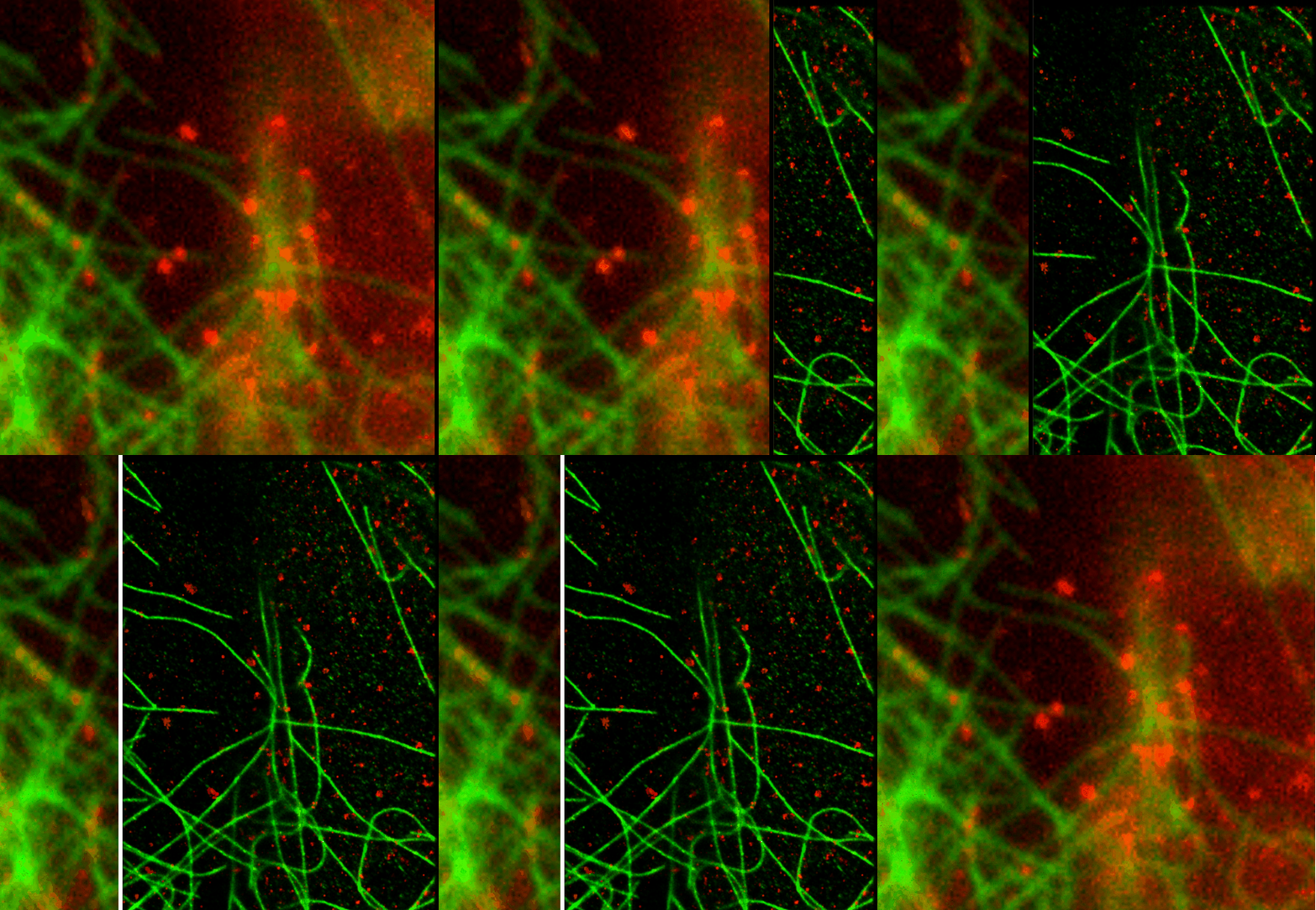
- Pointillism, STED, ExM
- Quantification assistance
Intra-vital imaging
- Anesthesia system (murine)
- Surgery
- Contrast SHG / THG
- Murine, fish, ovo models
- Biosafety Level 2 BSL2


Images processing
Cochin Image Database
- Online image backup and archiving
- Online processing
- Image restoration by 3D deconvolution
- Transfer
- Sharing
- Data: light microscopy, electron microscopy, ultrasound, cytometry, histology, genomics, proteomics


