

Team members


Projet
Expansion microscopy (ExM) is a sample preparation technique that enables the identification of structures at the nanometer scale. Cells or tissues are embedded in an acrylamide/bisacrylamide gel, which is then swollen to enlarge the sample. The mechanical enlargement of the sample causes labeled proteins to spread apart, making two objects that were previously optically indistinguishable now identifiable.
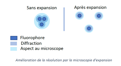
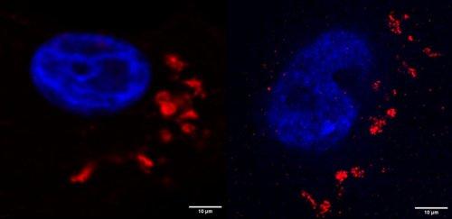
Several protocols are possible; the simplest one allows the sample to be magnified 4.5 times (proExM). More recent protocols make it possible to magnify the sample up to 20 times (iExM). ExM is a technique that bridges the gap between conventional microscopy and more advanced super-resolution techniques such as STED or dSTORM.
We are developing this technique on our platform so that we can offer it as a “turnkey” solution to everyone.
Offres stage
networks