Dehydration, impregnation and embedding in paraffin blocks
The HISTIM facility handles all types of organs and all species. After collection and fixation of the tissues by the user, the samples have to be brought in ethanol 70% on the platform with the Excel form (in French) «Demande d'inclusion coupe paraffine», in paper version. The paraffin dehydration and impregnation automaton is run on Tuesdays and Thursdays at 4pm. Please bring your samples on time. The paraffin block inclusion is realized the next day and the recoverable blocks in the afternoon.


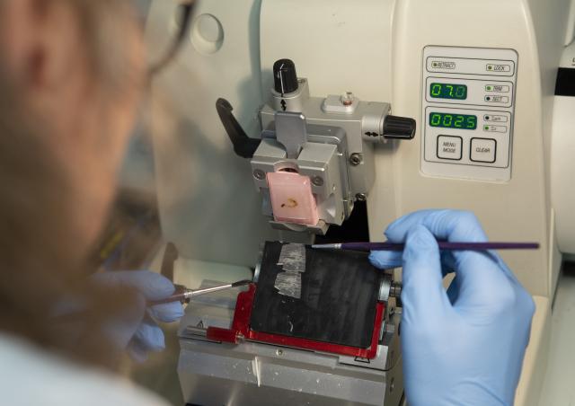
Tissue Sectioning on microtome or cryostat
The platform makes section from paraffin blocks on microtome and on cryostat from frozen tissue. Sections are used for various applications:
- On slides, for histological staining, immunostaining, HIS, TUNEL, etc.
- On tubes, for extraction and analysis of biological molecules
The facility is able to treat different types of blocks and special tissues, for example:
- Spheroids/Organoids
- Tissue Micro Array (TMA) block
- Section of cells (aggregates included in agarose, isolated pancreatic ilots)
- Decalcified hard tissue
The Excel form (in French) "Demande d'inclusion coupe paraffine" (if possible, link to the file provided in the appendix) must be completed and attached in the "Open Iris" request.
The Excel form (in French) “Demande coupe cryostat» (if possible, link to the file provided in the appendix) must be completed and attached in the "Open Iris" request.
Standard and specific histological staining
The platform provides standard morphological stainings to highlight the complete cellular and structural morphology and organization of tissues, on paraffin sections or frozen tissues. Structural abnormalities, inflammation and cellular infiltration, tumours, pathological signs (steatosis, fibrosis, etc.) can be identified.
Here are some examples of staining:
- Hematoxylin/Eosin (staining of nuclei in blue and and cytoplasms in pink)
- Hematoxylin/Erythrosin/Saffron (yellow fibres)
- Masson’s Trichrom (blue fibres)

It is also possible to highlight specific structures and components (not exhaustive):
- Sirius Red: Collagen and Fibrosis
- Pearls: Iron
- Oil-Red-O: Lipid droplets and steatosis
- PAS (Periodic Acid Schiff): Mucopolysaccharids
- Alcian Blue: Acid Mucopolysaccharids
- May- Grunwald-Giemsa: immune cells
- Gram: bacteria
- Alizarin Red: Calcium


Immunostaining
Tissue characterization is also performed at a molecular level with the identification of target proteins, by different types of Immunostaining:
- Single Fluorescence
- Single Chromogenic (DAB/ Brown or Red)
- Double or triple fluorescence staining
- Double brightfield immunostaining ( Brown and one other chromogenic)
- OPAL Technology Multiplexing (In development)
- Ultivue Technology Multiplexing (In development)
Immunolabeling is performed on an automaton, Leica Bond RX. Users provide primary antibodies and the platform provides all the other reagents and secondary antibodies:
- Fluorescence: anti-rabbit or anti-souri, Dye light 488 green or Dye light 555 red
- Brightfield : HRP-coupled secondary antibodies: anti-rabbit, mouse, rat or goat; ALP-coupled secondary antibodies: anti-rabbit, mouse.
The Excel form (In French) « Demande d’immunomarquage» (if possible, link to the file provided in the appendix) must be completed and attached in the "Open Iris" website.


Production of TMA (Tissue Micro Array) blocks
The HISTIM facility owns a Tissue Micro Arrayer. The production of a TMA block consists of collecting tissue cores from dozens of donor blocks on a single recipient paraffin block. This reduces the number of blocks to be processed for:
- Saving time: fewer blocks, fewer slides to process
- Cost savings: fewer reagents to use
- Improved reliability of results: all samples are processed at the same time under identical experimental conditions.
- Better analysis: all samples are analyzed in one run on the same slide.
The Excel form (in French) «Demande de blocs Tissue Micro Array (TMA)» (if possible, link to the file provided in the appendix) must be completed and attached in the « Open Iris » website.
Virtual slides production with high speed slide scanner
The platform has a slide scanner, the "Lamina slide scanner" (250 automatic slides) from Akoya Perkin Elmer. This automaton is able to acquire your entire slides in few minutes, whether in brightfield (histological stainings and brightfield immunostaining) or fluorescence, and to creat high-resolution virtual slides. Virtual slides are then very easily observable with specific software, such as Case Viewer or Pannoramic Viewer, which can be downloaded free of charge from the internet. With these softwares called «virtual microscopes», you can zoom in on the regions of interest without losing any resolution, make captures, annotations, measurements, manual counts.
The platform scan your slides, at your request, every Monday and Tuesday morning.
The Excel form (in French) « Demande d’acquisition de lames virtuelles» (if possible, link to the file provided in the appendix) must be completed and attached in the "Open Iris" website:
- In the service request “paraffin service request” when the slides have been performed on the facility on FFPE tissue
- In the service request “frozen service request” when the slides have been performed on the platform from frozen tissues.
- In the Service Request “Slides Only: Virtual Slide Scan Request” when you bring your own slides, already stained or immunostained.


Image analysis
Depending on the project, the type of analysis, the degree of analysis requested and after discussion, the platform can support all or part of the image analysis.
The platform can also help you with observation and analysis of the slides.
It can also train you in the INFORM software to perform autonomous analysis.
With the INFORM software, analyies can be done on histological staining, immunofluorescence or visible immunolabelling, for different applications:
- Tissue segmentation
- Percentage of stained area Quantification
- Percentage of positive and negative cells Quantification
- Cell Phenotyping.